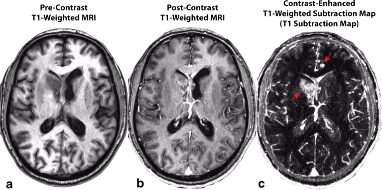
Fig. 1.
Construction of contrast enhanced T1-weighted subtraction maps in a recurrent glioblastoma patient treated with bevacizumab. A) Pre-contrast T1-weighted MR image. B) Post-contrast T1-weighted MR image. C) T1 subtraction map calculated by voxel-wise subtraction of pre-contrast from post-contrast T1-weighted images highlighting areas of increased contrast enhancement. Red arrows show two subtly enhancing lesions that are easily identified on T1 subtraction maps

