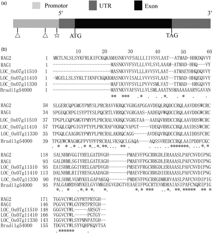
Figure 1.

Structural and sequence analysis of RAG2. (a) A schematic representation of the exon and intron organization of RAG2. RAG2 consists of one exon (black box) with an 82‐bp 5′UTR (grey box) and a 200‐bp 3′UTR (grey box). Two ATGCAAAA (triangle, −1028 bp, −252 bp) and one CTTTAGTCTT (pentagon, −21 bp) cis‐element in RAG2 promoter region. (b) Protein sequence alignment of RAG2 with RAG1, LOC_Os07g11510, LOC_Os07g11410, LOC_Os07g11330 and Bradi1g54000. Residues marked with asterisks and dots are highly conserved and semiconserved, respectively. A dash ‘–’ denotes a gap in the alignment.
