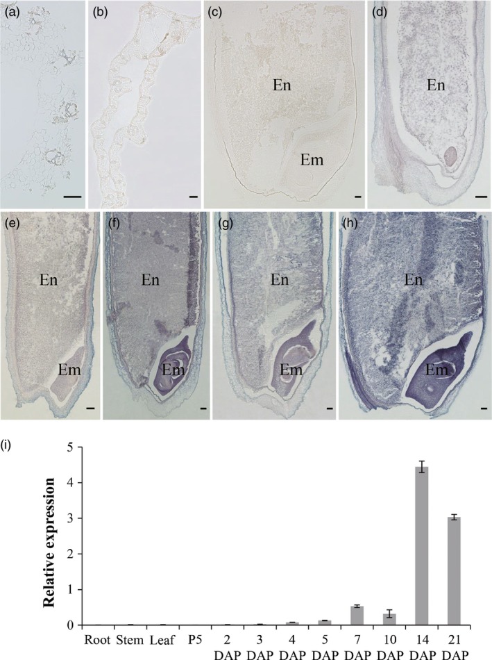
Figure 2.

Spatial and temporal expression pattern of RAG2. (a–h) RNA in situ hybridization of RAG2. (a) Stem. (b) Leaf. (c) Sense. (d) 3‐d seed. (e) 5‐d seed. (f) 7‐d seed. (g) 10‐d seed. (h) 14‐d seed. En, Endosperm; Em, Embryo. Bar: 100 μm (a–h). (i) qRT‐PCR analysis of RAG2. Total RNAs were extracted from root (booting stages), stem (booting stages), leaf (1 day before heading), P5 is panicle at stage 5 (pollen mother cell formation stage), developing seeds (2, 3, 4, 5, 7, 10, 14 and 21 DAP). Data are mean ± SE for three replicates.
