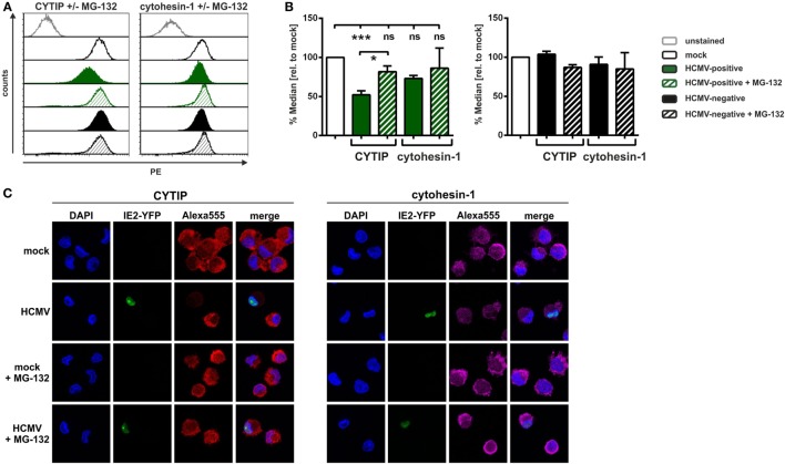
Figure 6.
In human cytomegalovirus (HCMV)-infected mature DCs (mDCs) CYTIP is degraded via the proteasome. Mature dendritic cells were mock- or HCMV-infected, treated with or without MG-132 and harvested 24 hpi. (A,B) Subsequently, intracellular staining with antibodies specific for CYTIP or cytohesin-1 was performed, and samples were subjected to flow cytometric analyses. (A) One representative experiment is shown for CYTIP (left panel) or cytohesin-1 (right panel) levels. Mock conditions (black lined histograms), HCMV-positive cells (green filled histograms), and HCMV-negative cells (black filled histograms) are shown, while the respective MG-132-treated samples are depicted as striped histograms. (B) Summarized data of at least three independent experiments of CYTIP and cytohesin-1 protein levels, analyzed via intracellular flow cytometry. Median values of HCMV-positive mDCs with and without MG-132 treatment (green filled and green striped bars, respectively; left panel) as well as HCMV-negative mDCs with and without MG-132 treatment (black filled and black striped, respectively; right panel) were calculated relative to the respective mock conditions with and without MG-132 (white bars, set to 100%). Significant changes (*** = p < 0.001; * = p < 0.05) are marked by asterisks, non-significant changes (p > 0.05) as “ns.” (C) Immunofluorescence staining of CYTIP (left panel) and cytohesin-1 (right panel). The IE2-YFP fusion protein of the HCMV TB40E/IE2-EYFP strain allows direct determination of HCMV-positive and HCMV-negative cells. The nucleus was visualized using DAPI. The experiment was performed three times and representative data are shown.

