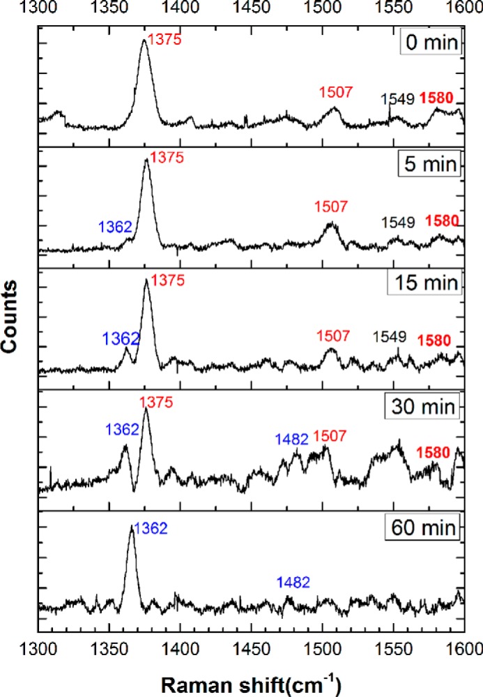
Figure 8.

Resonance Raman spectra of FeIII-Ngb treated with sulfide. High-energy region of the spectra monitoring the time-dependent changes in FeIII-Ngb (400 μm) in 100 mm HEPES buffer, pH 7.4, containing 20% glycerol (v/v) incubated with Na2S (10 mm) at 25 °C under aerobic. The data were obtained using 413.1-nm excitation with 10–25-milliwatt laser power. Measurements for each sample took ∼10 min (various exposure time and accumulations). Initially, the observed ν2, ν38, ν3, and ν4 vibrations at 1580, 1549, 1507, and 1375 cm−1, respectively, represent a 6-coordinate ferric low-spin heme. With time, the appearance of the ν3 and ν4 vibrational frequencies at 1482 and 1362 cm−1, respectively, indicates a 6-coordinate ferrous low-spin heme.
