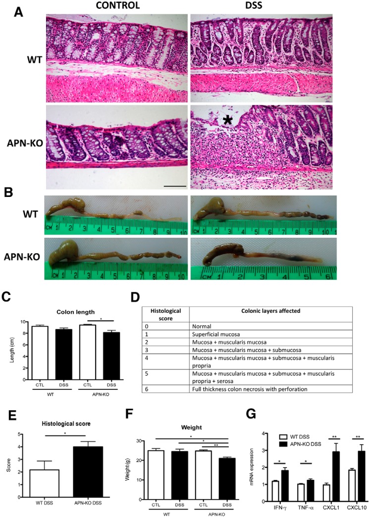
Figure 1.
APN-KO mice are more susceptible to DSS-colitis than WT mice. A, H&E histology from WT and APN-KO of the descending colon, asterisk (*) shows inflammation and architectural distortion. B, representative images of colonic shortening in APN-KO colitic mice, and C, colonic length reduction following DSS between all groups. D, a histological scoring system used to evaluate the mouse cohorts, and E, APN-KO mice had 2-fold more damage that WT. F, reduced weight after DSS treatment in APN-KO colitic mice compared with other groups. G, proinflammatory cytokine profile of APN-KO colitic mice compared with WT colitic mice, showing an increase in IFN-γ and TNF-α (p < 0.05 for both groups), CXCL1 and CXCL10 (p < 0.01 for both groups). Scale bar, 100 μm. *, p < 0.05, ** p < 0.01.

