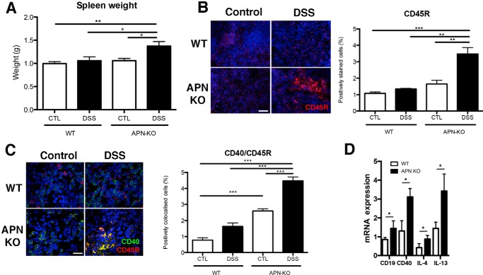
Figure 7.
Enhanced splenic B cell response in DSS-colitis in APN-KO mice. A, spleens of APN-KO DSS-treated mice were heavier than WT controls, DSS-treated groups, and APN-KO controls. Immunofluorescent analyses of the spleens confirmed that: B, CD45R staining (red) was increased in APN-KO DSS mice compared with controls and WT DSS groups (p < 0.01 and p < 0.0001). C, CD40 (green) and CD45R (red) colocalization was increased in APN-KO DSS versus WT DSS colons (p < 0.001). APN-KO DSS colons had increased CD40/CD45R staining versus WT and APN-KO control groups (p < 0.001 all comparisons). Scale bar, 25 μm. D, splenic inflammatory cytokine profile by qRT-PCR analysis in WT and APN-KO treated with DSS, shows increases in CD19 (p < 0.05), CD40 (p < 0.001), IL-4 (p < 0.05), and IL-13 (p < 0.05). *, p < 0.05, ** p < 0.01, *** p < 0.001.

