Figure 1.
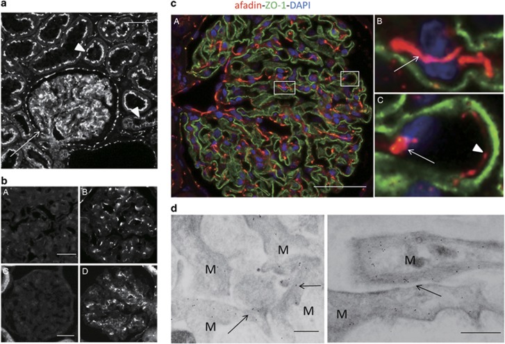
Expression of afadin in the kidney cortex. (a) Sections of human kidneys were stained with anti-afadin antibody. Afadin signals were detected in glomeruli (arrow), and apical side of tubular epithelial cells (arrowheads). Scale bar, 50 μm. (b) Sections of human (A and B) and rat (C and D) kidney were stained with anti-afadin antibody preadsorbed with anti-myc immunoprecipitates from cell lysates of HEK293 cells expressing myc-tagged l-afadin (A and C) or control vector (B and D). (c) Dual-labeling immunofluorescence of afadin (red) and ZO-1 (green) in the human kidney. Strong afadin signals were detected in the mesangium, particularly in cell–cell adhesions between mesangial cells (B, arrow) and between mesangial and endothelial cells (C, arrow). Afadin signals were also detected at cell–cell adhesions between endothelial cells of glomerular capillaries (C, arrowhead). Higher-magnification images corresponding to squares in A are shown in B and C. Scale bar, 50 μm. (d) Immunogold staining of afadin detected using immunoelectron microscopy. Immunogold particles specific for afadin were detected in the mesangial cell–cell adhesion sites (arrows). Scale bars, 200 nm. M, mesangial cell.

