Figure 5.
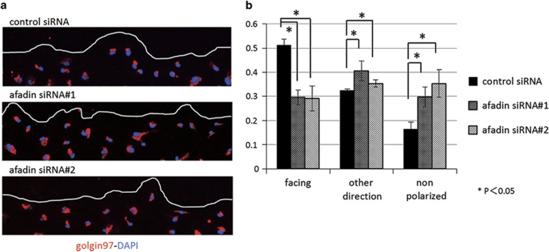
Impaired formation of front-rear polarity in afadin-depleted mesangial cells. (a) Confluent human mesangial cell monolayers of control siRNA and siRNAs #1 and #2 for afadin were manually scratched and cultured for 24 h. Cells were stained with goldin97 (golgi complex marker, red) and DAPI (blue). The line indicates the leading edge of the wound. (b) The percentage of Golgi apparatus facing the wound, facing the other direction, and non-polarized with respect to the wound was calculated as described in the Materials and Methods. *P<0.05 using Student's t-test. Data are shown as the mean±s.d. of three independent experiments.

