Figure 4.
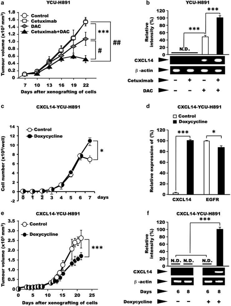
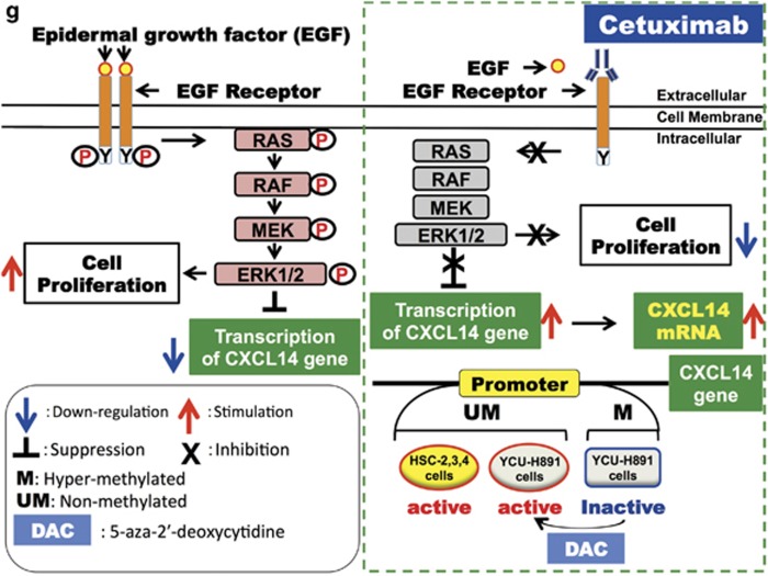
Effects of cetuximab and/or DAC, or ectopic expression of CXCL14 on tumour growth and CXCL14 mRNA levels in YCU-H891 cells. (a, b) BALB/c nude mice (24 females, 5-week old) were subcutaneously inoculated on the dorsal side with YCU-H891 cells (1 × 10⁷ per site). Seven days after inoculation (at a tumour size of ~100 mm3), the animals were randomly divided into four groups and intraperitoneally administered Dulbecco's phosphate-buffered saline (control), cetuximab (cetuximab-only, 10 mg/kg), DAC (DAC-only, 5.0 mg/kg), or both cetuximab (10 mg/kg) and DAC (5.0 mg/kg; cetuximab+DAC) three times per week. Tumour volumes were measured once every 3 days by a person different from the one who injected the reagents (a) and were calculated with the formula, (a × b × b)/2, where ‘a' is the long diameter and ‘b' is its short diameter of the tumours. Total RNA was isolated, and the expression levels of CXCL14 mRNA were determined as described in the legend for Figure 1d. To compare the expression levels of CXCL14 between the DAC-only and cetuximab+DAC groups, we quantified the level of CXCL14 mRNA in each group by using quantitative PCR (qPCR; b). One of the representative data set of two similar experiments is presented. (c, d) In another series of experiments, we engineered YCU-H891 cells to ectopically express CXCL14 under the control of doxycycline (CXCL14-YCU-H891 cells). The cells (1 × 10⁴ per well) were plated into 24-well plates (Sumitomo Bakelite, Osaka, Japan) in DMEM medium containing 10% fetal bovine serum, and the next day half of the plates were treated with 0.2 μg/ml of doxycycline (Doxycycline, Takara). The experiments were repeated three times and yielded similar results. The number of cells was counted using a Coulter Z1 Counter (Coulter Electronics Ltd, London, UK; c). One day after the cells had been plated (5 × 10 × 10 × 10 × 10) into six-well plates, half of the plates were treated with 2 μg/ml doxycycline. After 24 h, RNA was isolated and qPCR was performed as described in the legend for Figure 3f and g, and relative expression levels of mRNAs of CXCL14 and EGFR were determined (d). (e, f) For the in vivo experiment, the cells (1 × 10⁷ per site) were subcutaneously injected into the backs of BALB/c nude mice (12 female, 5-weeks old). Seven days after tumour cell inoculation (at a tumour size of ~100 mm3), the mice were randomly divided into two groups. One group was fed a 5% (w/v) sucrose solution containing 2 mg/ml doxycycline (Takara), whereas the other group was fed a 5% sucrose solution (control). The tumour sizes were measured five times per week, as described above (e). To confirm the enhanced expression of CXCL14 in the presence and absence of doxycycline, we removed tumour tissue before (day 6) and after doxycycline administration (day 8), extracted the total RNA from the tissue samples and measured the levels of CXCL14 mRNA (f), as described above. The values are expressed as the mean±s.d. (n=6). ***P<0.001, #P<10−4, ##P<10−5 (Student's t-test). A schematic representation of the effects of EGF and cetuximab on cell proliferation and the expression of CXCL14 is presented in g.


