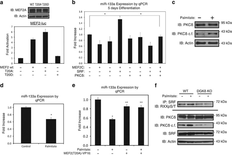
Figure 3.
Mutational analysis of threonine-20. (a) HEK293 cells were transfected with MEF2A (MEF2 wt), or a plasmid containing MEF2A where threonine-20 is mutated to a neutral alanine (T20A) or phospho-mimetic aspartic acid (T20D), as indicated, and subjected to western blot (above). 10T1/2 cells were transfected, as above, along with MEF2-driven luciferase reporter gene (MEF2-luc). Extracts were subjected to luciferase assay, where β-galactosidase assay was used to correct for transfection efficiency (below). All assays were done in triplicate. (b) C2C12 myoblasts were transfected with MEF2C, SRF, or PKCδ, as indicated. Following recovery, cells were differentiated in low serum media for 5 days, harvested for RNA, subjected to qPCR assay. (c and d) Following 5 days of differentiation, C2C12 myotubes were treated with 200 μM palmitate conjugated to 2% albumin in low glucose media overnight. Control cells were treated with 2% albumin alone. Myotubes were harvested for RNA or protein, and assayed by immunoblot (c) or qPCR (d). (e) C2C12 myoblasts were transfected with MEF2-VP16 fusion where threonine-20 is mutated to a neutral alanine [MEF2(T20A)-VP16], or control plasmid. Cell was differentiated for 5 days and treated with 200 μM palmitate, as indicated. Myotubes were harvested for RNA and assayed by qPCR. (f) Wild-type (WT) or diacylglycerol kinase-δ knock-out (DGKδ KO) embryonic fibroblasts treated with palmitate, as described above. Extracts were immunoprecipitated (IP) with SRF antibody and probed using an antibody that recognizes phospho-serines/threonines with arginines at the –3 position (RXXpS/T) or immunoblotted (IB), as indicated. PKCδ=catalytic fragment of PKCδ. Data are represented as mean±S.E.M. *P<0.05 compared with control. **P<0.05 compared with palmitate treatment

