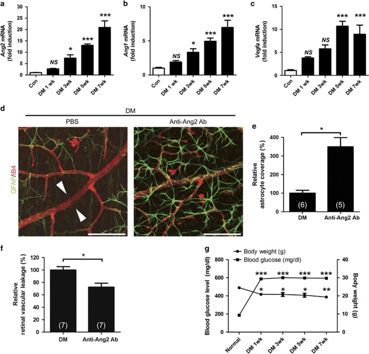
Figure 2.
Inhibition of Ang2 reduces the astrocyte loss and vascular leakage in early diabetic retina. (a–c) Retinal mRNA was determined in 1, 3, 5, and 7 weeks from streptozotocin-induced diabetic mice (DM) and control mice (Con) retinas by qPCR, and normalized to Rn18s mRNA. (a) Ang2 mRNA. (b) Ang1 mRNA. (c) Vegfa mRNA. (d–f) Anti-Ang2-neutralizing antibody (Anti-Ang2 Ab, 1 μg) or PBS was intravitreally injected to 2-week-old DM. Retinal astrocyte and retinal vascular leakage were evaluated 1 week after the injection in 3-week-old DM. (d) Focal astrocyte loss shown in diabetic retina (PBS) is rescued in anti-Ang2 Ab-injected diabetic mice (original magnification × 400; scale bar, 100 μm). White arrowheads indicate loss of astrocytes on diabetic retinal vessels. (e) Relative astrocyte coverage (% Anti-Ang2 Ab) is calculated by colocalized area per IB4+ vascular area and normalized to the value of control mice. (f) Relative retinal vascular leakage (% Anti-Ang2 Ab) with FITC-dextran is shown normalized to the value of control mice. (g) Body weight and blood glucose level of the STZ-induced diabetic and non-diabetic control mice at 7 weeks. The sample size for each group is indicated on the bar graph. The bar graphs represent mean±S.E.M.; *P<0.05, **P<0.01, and ***P<0.001 by ANOVA and Student's t-test

