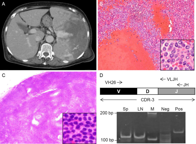
Figure 1.
Splenic bleeding of splenic marginal zone lymphoma (SMZL). (A) A dynamic CT scan of the abdomen on admission. (B) Histology of the excised spleen. Hematoxylin and Eosin (H&E) staining. Magnification, ×100, inset×1,000. (C) Histology of the excised lymph node. H&E staining. Magnification, ×20. Inset, ×400. (D) PCR amplification of complementarity determining region-3 (CDR-3). The spleen (Sp) and lymph node (LN) lanes show monoclonal bands of the same size. VH26, forward primer. VLJH, reverse primer for second PCR. JH, reverse primer for first PCR. M, size marker. Neg, negative control. Pos, positive control.

