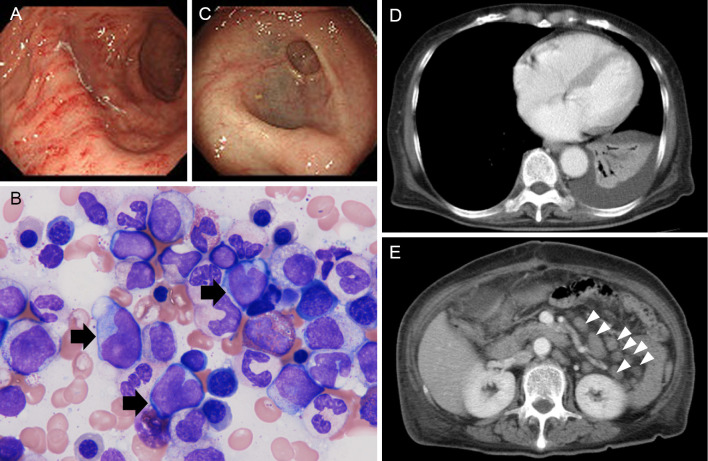
Figure 2.
Staging of SMZL. (A) Gastroendoscopy image. Mucosal biopsy revealed lymphoma invasion (data not shown). (B) A bone marrow smear. The black arrows show lymphoma cells. Wright-Giemsa staining. Magnification, ×1,000. (C) Colonoscopy image. The mucosae of the sigmoid colon and rectum were mildly opaque. (D) A contrast-enhanced CT scan of the lungs. Pleural effusion and atelectasis of the left lower lung are shown. (E) A contrast-enhanced CT scan of the abdomen. Arrowheads, swollen lymph nodes.

