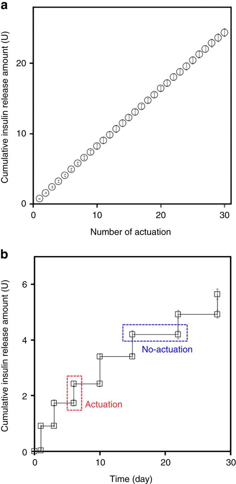
Figure 2. In vitro insulin release profiles of the MDP.
The MDP was fully immersed in pH 7.4 PBS and incubated in an incubator at 37 °C while being continuously shaken at 50 r.p.m. At each actuation, the external magnet device was applied at a constant distance from the top of the MDP by placing a 1 mm-thick glass slide between the external magnet device and MDP to simulate the presence of tissue after implantation, and removed almost instantaneously (<1 s) while the MDP was fully submerged in the release medium. An aliquot of the release medium was sampled at scheduled times and analysed by high-performance liquid chromatography. Details on this procedure are described in the Methods section. (a) Thirty consecutive actuations were applied at intervals of 10 min, and an aliquot was sampled after each of the actuations. With five distinct MDPs tested herein, the released amount of insulin was highly reproducible and was 0.81±0.04 U per actuation. Error bars are s.d. (b) The MDP was actuated at predetermined times of 1, 3, 6, 10, 15, 22 and 28 days while fully immersed in PBS for 28 days. Aliquots were sampled immediately before and after each of the actuations. With four distinct MDPs tested herein, there was almost no release of insulin during the period of no actuation. The released amount of insulin was also highly reproducible and was 0.80±0.09 U per actuation. Error bars are s.d.

