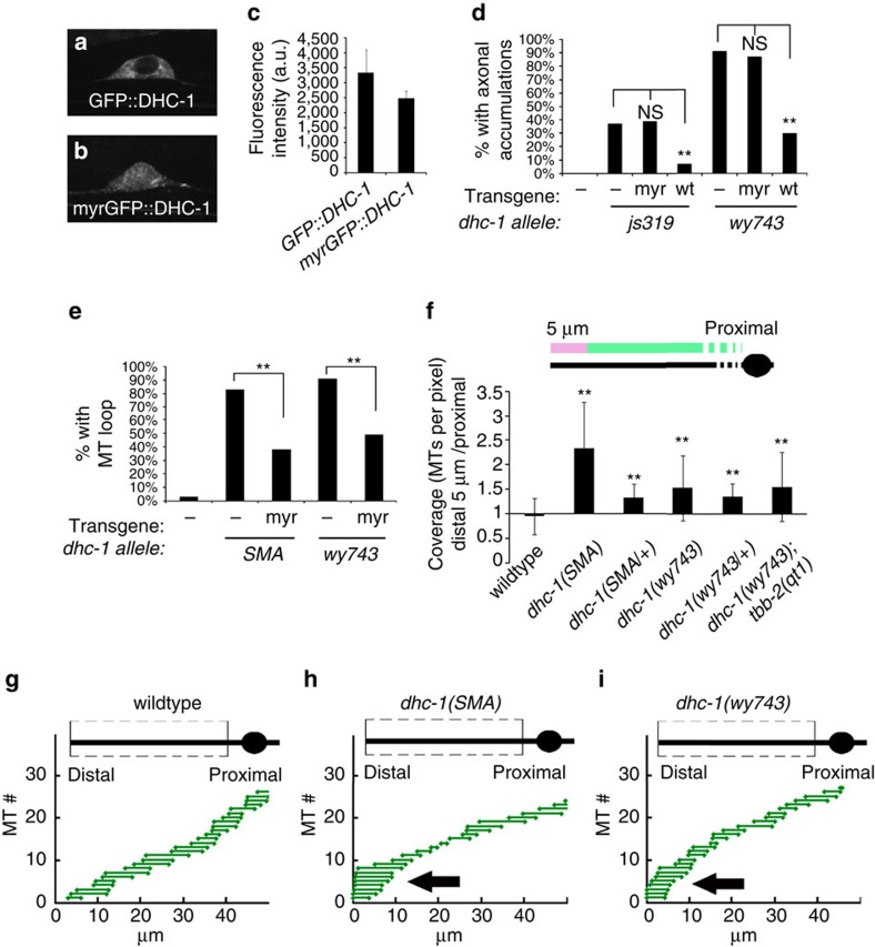
Figure 4. dhc-1 cortical rescue and a distal shift in MT distribution in dhc-1 mutants.
(a) GFP::DHC-1 is localized to the cytoplasm. (b) myrGFP::DHC-1 shows a distribution consistent with membrane localization. (c) Expression levels of GFP::DHC-1 and myrGFP::DHC-1 were quantified after imaging in identical settings. n=17 per genotype. (d) The myrGFP::DHC-1 construct does not rescue the accumulation of axonal SVPs (GFP::RAB-3). n=20–25, **P<0.05. (e) membrane tethered myrGFP::DHC-1 rescues MT loop formation in dhc-1 mutants. n=28–45, **P<0.05. (f) Ratio between MT coverage (number of MTs per pixel) in the distal 5 μm of the dendrite to the rest of the dendrite. The results indicate that the distribution of MTs is shifted distally in dhc-1 mutants. (g–i) Representations of MTs in the dendrites of indicated phenotypes. The representation is derived from a quantized GFP::TBA signal, where steps up indicate a MT start and steps down a MT end. MT ends were randomly assigned to the longest MT in the bundle for representation purposes. See Methods for details.

