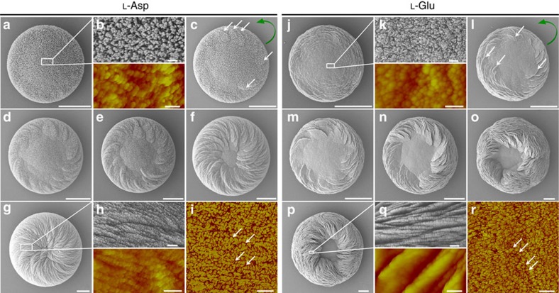
Figure 5. Growth evolution of chiral vaterite toroids grown in L-Asp or L-Glu.
SEM and AFM (coloured) images of toroid growth for 2 h (a,b), 3 h (b), 5 h (d), 8 h (e), 12 h (f) and 24 h (g,h) in the presence of L-Asp, and for 1.5 h (j,k), 2 h (l), 3 h (m), 5 h (n), 10 h (o) and 24 h (p,q) in the presence of L-Glu. Oriented vaterite platelets (white arrows) emerge within several hours (c,l) at the outer edge of a flat substrate vaterite disc to begin the formation of a toroid with a counterclockwise chirality (green curved arrow), and these and other platelets continue spiral growth to encroach upon, and then obscure (in the case of Asp), the centrally located achiral vaterite core region. SEM images (upper panels of b,h,k,q) and AFM height images (lower panels of b,h,k,q) show that all vaterite elements in the toroids have nanoparticle substructure. AFM phase mode (i,r) shows inorganic vaterite nanoparticles (yellow) surrounded by organic amino acid (red) in the platelets. Scale bars, 4 μm (a,c–g), 200 nm (b,i,k,r), 400 nm (h), 10 μm (j,l–p), 400 nm (upper panel of q) and 200 nm (lower panel of q).

