Fig. 5.

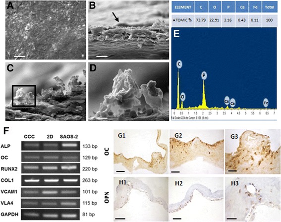
Osteogenic differentiation on Collagen I-based Cell Carrier (CCC). a, b SEM images of differentiated DPPSC adhered on CCC surface (black arrow). c SEM image of differentiated DPPSC with hydroxyapatite deposition on CCC surface. d SEM image of hydroxyapatite deposition on CCC. Scale bars: 100 μm (a), 20 μm (b), 10 μm (c), 2 μm (d). e Microanalysis of the CCC surface with atomic concentrations. f RT-PCR gene expression analysis of differentiation markers (OC, ALP, COL1) and adhesion markers (VCAM1, VLA4) in DPPSC cultured on CCC, DPPSC cultured on 2D (plastic surface) and SAOS-2 cultured on CCC. GAPDH was used as a housekeeping. g, h Immunohistochemistry of differentiation markers (OC, OPN) in differentiated DPPSC on CCC. Scale bars: 1000 μm (g1, h1), 400 μm (g2, h2); 200 μm (g3, h3)
