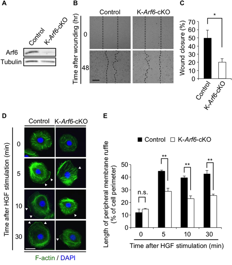
Figure 5. HGF-stimulated cell migration and peripheral membrane ruffle formation are impaired in Arf6-deleted keratinocytes.
(A) Ablation of Arf6 from keratinocytes. Lysates of primary cultured keratinocytes prepared from control and K-Arf6-cKO mice were immunoblotted with anti-Arf6 and anti-tubulin antibodies. (B,C) Scratch-wound assay of primary cultured keratinocytes treated with 50 ng/ml of HGF. Representative images (B) and quantitative data of wound closure at 48 hr after scratching (C) were shown. (D,E) Primary cultured keratinocytes prepared from control and K-Arf6-cKO mice were stimulated with 50 ng/ml of HGF for the indicated time, and immunostained with F-actin (green) and DAPI (blue). Shown are representative images (D) and quantitative data of the peripheral membrane ruffle formation (percentage of peripheral membrane ruffle length to cell perimeter) of 50 cells (E) from three independent experiments. Allow heads indicate the region of peripheral membrane ruffles. Data show the mean ± SEM of five (C) and three independent experiments (E). Statistical significance was calculated using Student’s t-test; n.s., not significant, *P < 0.05, **P < 0.01, Scale bar, 200 μm (B), 10 μm (D).

