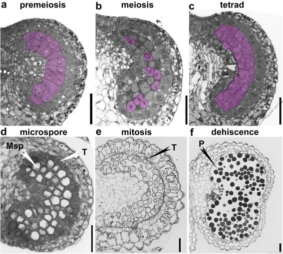
Fig. 2.

Histological characterization of anther development in tobacco (Nicotiana Benthamiana). Semi-thin transverse sections of tobacco anthers at pre-meiosis (a), meiosis (b), tetrad (c), microspore (d), mitosis (e), and dehiscence (f) stages. Pink-shaded cells in (a–c) are sporogeneous cells (a), pollen mother cells (PMC) at metaphase (b), or tetrads (c). Areas highlighted by the dotted line in (b) are a cluster of PMCs. Msp, microspores; P, pollen; T, tapetum. Bars, 50 μm
