Fig. 1.

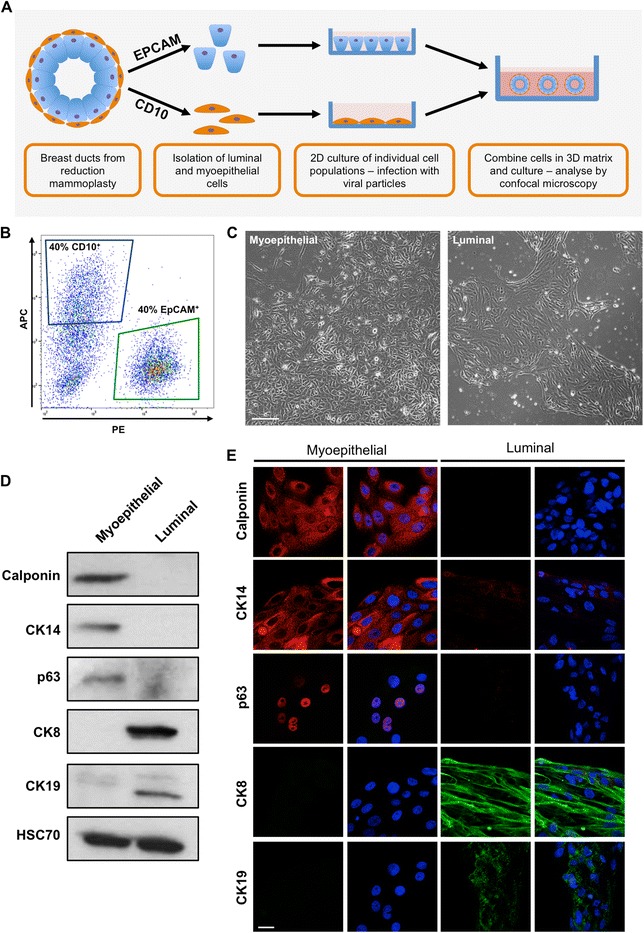
Isolated myoepithelial and luminal cells maintain their characteristics in vitro. a Schematic of proposed ductal model. b Representative fluorescence-activated cell sorting plots of reduction mammoplasty specimens separated by expression of CD10 (allophycocyanin fluorescence, blue gate) and epithelial cell adhesion molecule (EpCAM; phycoerythrin fluorescence, green gate). c Representative light micrographs of isolated myoepithelial and luminal cells grown in vitro for 10 days. Images taken at × 4 original magnification. Scale bar = 100 μm. d and e Western blot (d) and confocal (e) analysis of calponin, p63, cytokeratin (CK) 14, CK8 and CK19 expression in myoepithelial and luminal cells grown for 10 days in culture. Cell nuclei are labelled with 4′,6-diamidino-2-phenylindole (blue). Images and plots are representative of cells derived from at least three donors. Scale bar = 20 μm
