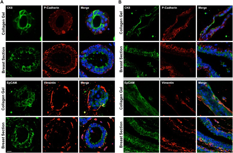
Fig. 3.

Spheroid and ductal structures formed in collagen gels recapitulate a physiological breast bilayer. Expression of cytokeratin (CK) 8, P-cadherin, epithelial cell adhesion molecule (EpCAM) and vimentin spheroid (a) and ductal (b) structures formed from myoepithelial and luminal cells grown in collagen for 21 days. Representative images from sections of normal human breast ducts (a and b) are also presented. Cell nuclei are labelled with 4′,6-diamidino-2-phenylindole (blue). Scale bar = 20 μm. Images are representative of structures formed from cells derived from at least three donors
