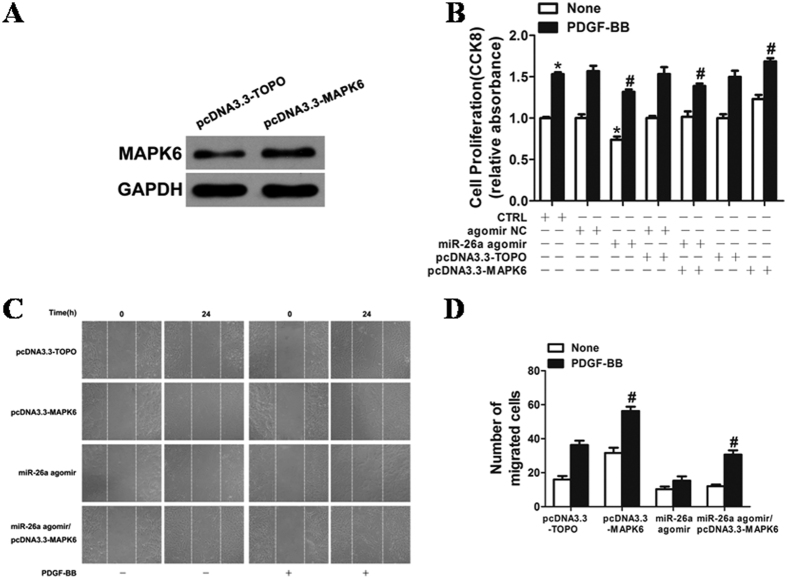
Figure 6. Overexpression of MAPK6 markedly augmented VSMC proliferation and wound closure after stimulation with PDGF-BB.
(A) VSMCs transfected with pcDNA3.3-MAPK6 significantly increased the expression of MAPK6. MAPK6 expression was determined by western blotting in VSMCs 48 h after transfection. The original images for (A) can be seen in Supplementary Fig. 3B–D. Overexpression of MAPK6 markedly augmented the VSMC proliferation and wound closure after stimulation with PDGF-BB. (B) VSMCs transfected with either miR-26a agomir or agomir NC (50 nM) were stimulated with or without PDGF-BB (20 ng/ml) for 24 h, and cell proliferation was measured by the CCK8 assay (n = 6). *P < 0.05 represents statistical significance compared with agomir NC without PDGF-BB. #P < 0.05 represents statistical significance compared with miR-26a agomir with PDGF-BB. (C) VSMCs transfected with oligos were starved, and cell migration was measured after PDGF-BB stimulation for 24 h by the scratch wound assay. (D) Migrated cells were quantitated and are shown as the mean ± SD of the number of migrated cells from three independent experiments. *P < 0.05 represents statistical significance compared with the control.

