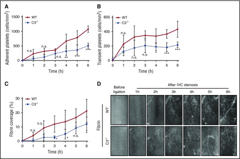
Figure 3.
Reduced fibrin formation in C3−/− mice in venous thrombosis. Adherent (A) and transient (B) platelets, and fibrin coverage (C) were compared between C3−/− mice and WT controls under IVC stenosis condition and imaged for over 6 hours using intravital microscopy (D) (n = 4-5; 3-4 visual fields per mouse; mean ± SD). Scale bar: 100 μm. Consecutive measurements were evaluated by 2-way ANOVA followed by Bonferroni posttest. *P < .05; **P < .01; ***P < .001. h, hour; n.s., not significant.

