
Figure 5.
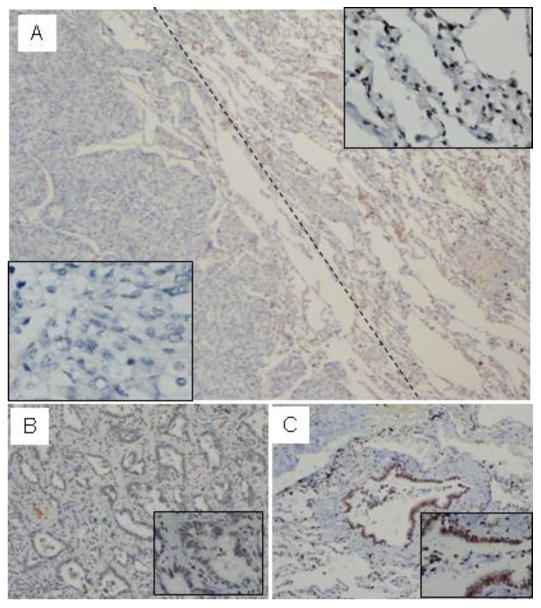
Menin expression is reduced in certain primary human lung cancer cells. Sections from paraffin-embedded lung cancer samples were stained with affinity-purified anti-menin antibody for immunohistochemistry staining. (A) Menin was easily detectable in the nucleus of the normal alveolar (Right) and in certain tumors (Left), where staining for menin was not detectable or markedly reduced (40×). (B) The menin staining was markedly reduced in lung adenocarcinoma, but in normal bronchiolo epithelial cells, the staining for menin was easily detectable, (C, 20×).