
Figure 2.
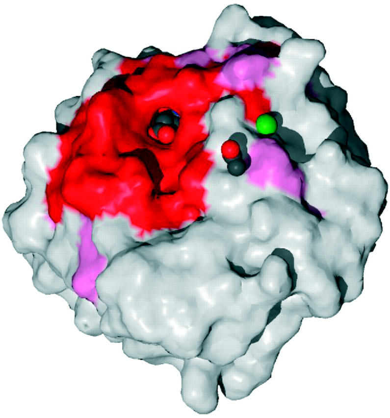
Surface of the glucosamine 6-phosphate synthase structure (PDB code 1gdo) coloured by residue conservation: red and pink for the most highly conserved regions, and blue for the most variable. The bound ligand—an l-glutamate—can be seen in spacefill representation within the highly conserved binding pocket. Also bound are an acetate ion and a sodium ion (green sphere).