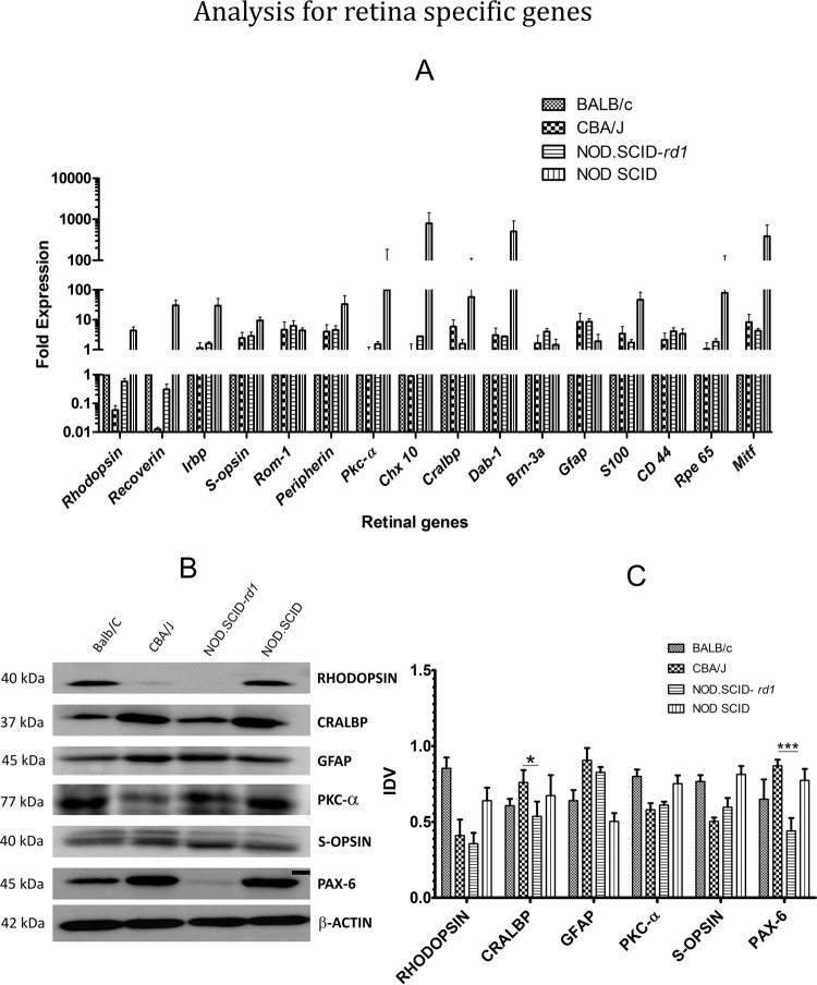
Fig. 3.
Analysis of retinal-cell-specific transcripts and protein levels. (A) Relative quantification of retinal cell transcripts in CBA/J and NOD.SCID-rd1 mice strain, compared with that of Balb/C at 4 weeks of age. The rod PR markers [Rho (Rhodopsin) and Rcvrn (Recoverin)] were highly downregulated in CBA/J, while NOD.SCID-rd1 showed a median expression level in between that of CBA/J and BALB/c. Irbp (interphotoreceptor retinoid-binding protein) expression remained similar in both CBA/J and NOD.SCID-rd1. Cone photoreceptor markers [S-opsin, Rom1, Prph (Peripherin)] remained comparable in CBA/J and NOD.SCID-rd1 and exhibited higher expression thanBALB/c. Retinal Müller glial cell markers (Gfap, S100 and Cd44) exhibited an upregulated expression in both CBA/J and NOD.SCID-rd1 as against BALB/c. RPE cell marker (Rpe65) remained unaffected while Mitf showed an elevated expression in both CBA/J and NOD.SCID-rd1. Amacrine cell marker (Cralbp) was slightly upregulated in NOD.SCID-rd1 and showed a highly elevated expression in CBA/J while Dab1 showed a comparable increase in both CBA/J and NOD.SCID-rd1 as against BALB/c. NOD.SCID-rd1 displayed an appreciative increase in the expression of retinal ganglion cell marker (Brn3a). CBA/J, however, indicated a marginal expression change as compared to BALB/c. Changes in the expression of retinal bipolar cell markers (Pkca and Chx10) were negligible in both CBA/J and NOD.SCID-rd1). (B) Representative images of western blot analysis for retinal proteins. (C) Integrated densitometry values (IDVs) indicated that CRALBP and PAX6 showed a lower expression in NOD.SCID-rd1 as compared to CBA/J, while both these strains exhibited slight downregulation in the expression of rod PR marker (Rhodopsin), cone PR marker (S-opsin) and bipolar cell marker (PKC-α) as compared to BALB/c. Moreover, the expression of GFAP was highly upregulated in both CBA/J and NOD.SCID-rd1 agreeing with the q-PCR results. ***P≤0.001 and *P≤0.05.

