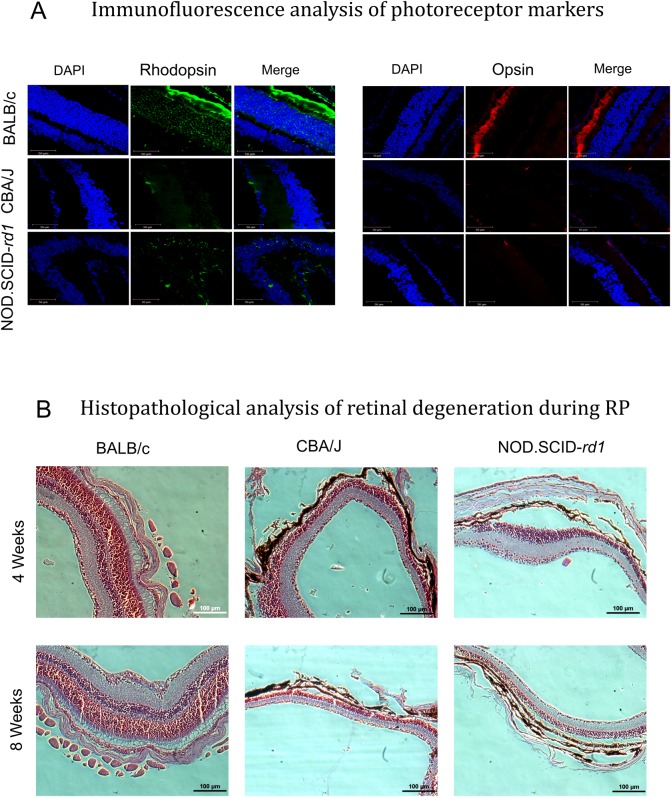
Fig. 4.
Immunofluorescence analysis of photoreceptor markers and histopathological changes during retinal degeneration. (A) Representative confocal micrographs (63×) for immunostaining of rod PR marker (Rhodopsin) and cone PR marker (S-opsin) in BALB/c, CBA/J and NOD.SCID-rd1 at 4-6 weeks of age. BALB/c strain with normal vision showed normal rod and cone PR staining in the outer segment of retina while rd1 model CBA/J had completely degenerated outer segments. NOD.SCID-rd1 also displayed outer segment degeneration, however, they had a few rhodopsin- and opsin-stained cells in the inner nuclear layer. The green color represents Rhodopsin-stained cells and red color represents S-opsin-stained cells in the retina. (B) Representative images illustrating histopathological analysis of retinal degeneration during RP in CBA/J and NOD.SCID-rd1 compared with BALB/c at 4 weeks and 8 weeks of age. The outer segment and outer nuclear layer had degenerated with detachment from RPE layer at various points in both CBA/J and NOD.SCID-rd1 by 4 weeks of age, with further thinning of inner nuclear layer by 8 weeks of age. BALB/c however retained intact retinal structure throughout.

