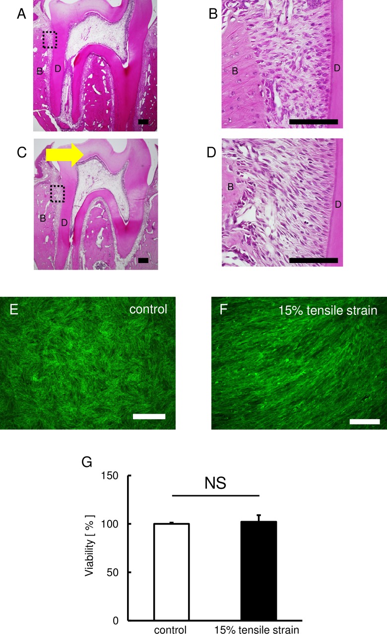
Fig. 1.
Effect of continuous tensile strain on the morphology of HPL cells. (A-D) Images of the PDL stained with H&E stain. Control group (A and B) and 5 days after tooth movement (C and D). B and D are higher magnification images of the boxed area in A and C. Arrow indicates the direction of tooth movement. B: bone, D: dentin. Scale bars: 100 μm. The effect of continuous tensile strain on cell morphology and cytoskeletal was assessed by means of F-actin staining. (E,F) The green fluorescence indicates the F-actin. Representative photographs of the control (E) and stretched HPL cells (F) are shown. Scale bars: 50 μm. (G) The effect of continuous tensile strain from the device on cell viability examined by using cell counting kit-8 (E). Percentage of controls is shown. Mean±s.d.; NS, not significant.

