Fig. 3.
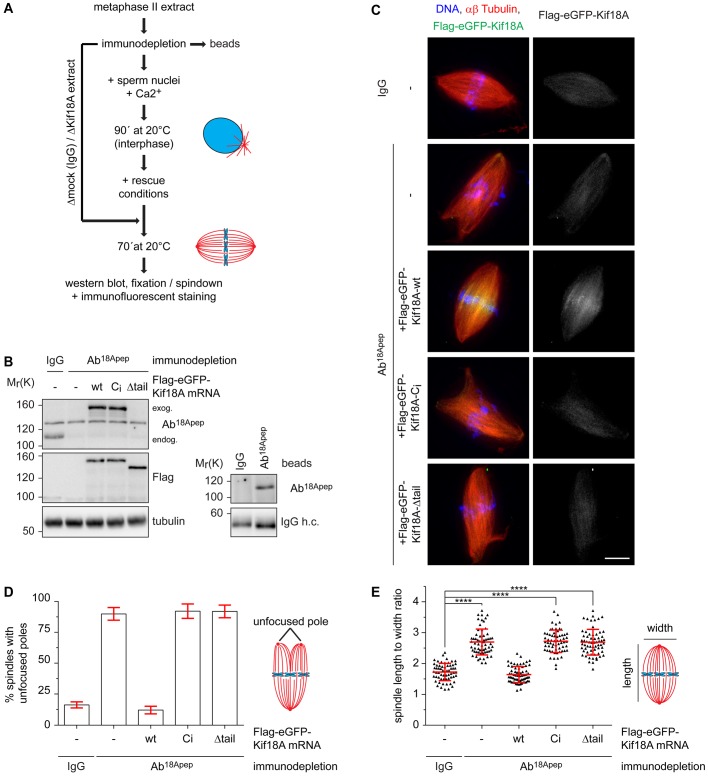
Xl_Kif18A is important for meiotic spindle structure. (A) Scheme of the depletion /add-back experiments. (B) Immunoblot of control (IgG)- or Kif18A (Ab18Apep)-depleted extracts supplemented with mRNA encoding wt, Ci or Δtail Flag-eGFP-Xl_Kif18A. Right panel shows immunoblot of IgG or Ab18Apep beads. (C) Representative fluorescence images of spindles obtained as described in A. DNA, αβ-tubulin, and Flag-eGFP-Xl_Kif18A are shown in blue, red and green, respectively. Scale bar: 10 µm. (D) Quantification of spindle length to width ratio. (E) Quantification of spindles with multiple/unfocused poles (more than 60 spindles analyzed per condition, mean±s.d., unpaired t-test: ****P≤0.0001).

