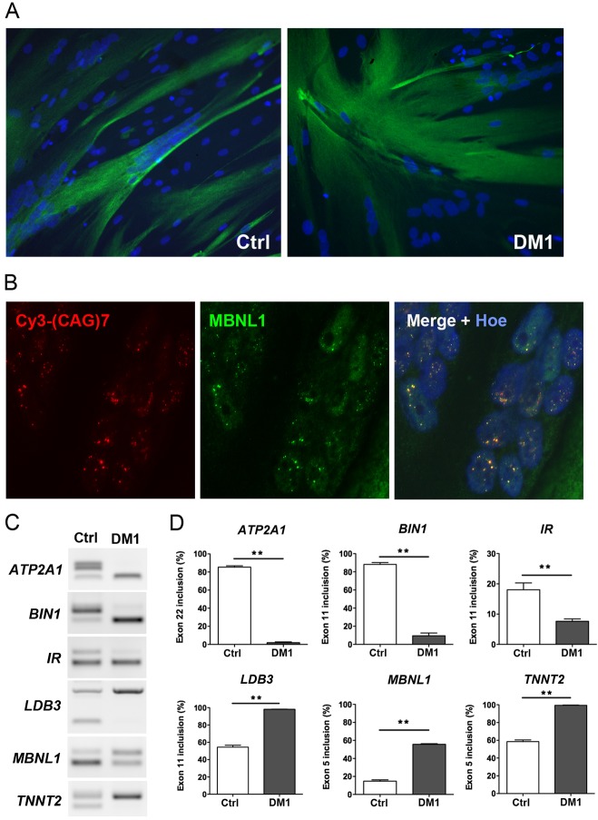
Fig. 3.
Characterization of DM1 and Ctrl myotube cultures derived from conditional MyoD-converted fibroblast cell lines. (A) Desmin (Desm) immunofluorescence staining (green) of myotube cultures derived from conditional MYOD1 converted Ctrl (left) and DM1 (right) fibroblast cell lines under permissive conditions to express MYOD1. (B) FISH using a Cy3-CAG7 probe (red) and MBNL1 immunostaining (green) showing the colocalization of MBNL1 with nuclear CUGexp-RNA aggregates in muscle converted DM1 immortalized fibroblasts. Hoe, Hoechst staining. (C,D) RT-PCR analysis and quantification of alternative splicing changes in ATP2A1, BIN1, IR, LDB3, MBNL1 and TNNT2 transcripts extracted from myotube cultures derived from converted DM1 and Ctrl fibroblast cell lines (n=4 for each condition). Data are expressed as mean±s.e.m. Comparison with Mann-Whitney test; **P<0.01.

