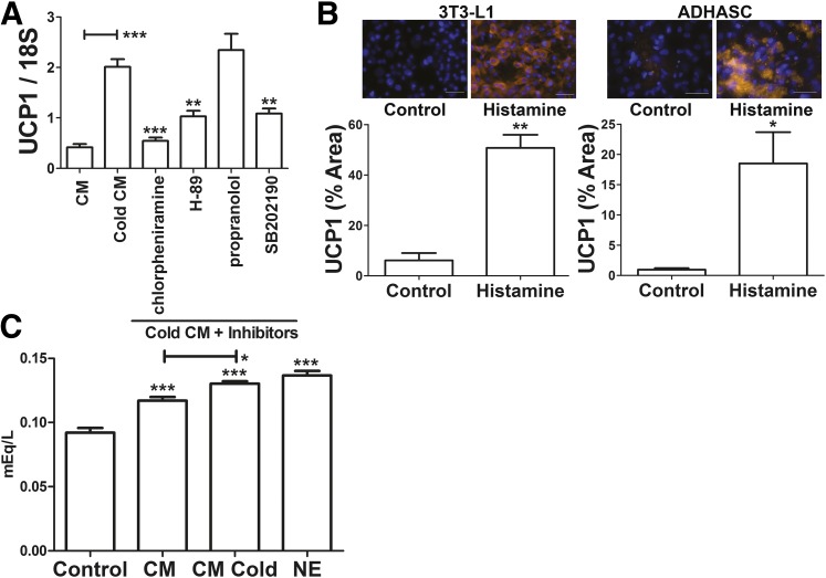
Figure 6.
Histamine in mast cell CM from cold-treated mast cells induces UCP1 expression in adipocytes through a PKA-dependent mechanism. A: Differentiated 3T3-L1 adipocytes were treated with vehicle (control), 5 μmol/L chlorpheniramine, 50 μmol/L H89, 10 μmol/L propranolol, or 100 nmol/L SB202190 in adipocyte differentiation media for 30 min as indicated. The adipocytes were then incubated with 50% mast cell CM (from cold-treated mast cells allowed to recover for 4 h at 37°C [cold CM]) and 50% adipocyte differentiation media containing the same final concentration of inhibitors or vehicle control for 4 h. The cells were harvested, and UCP1 mRNA expression was measured. Data are mean ± SEM (n = 3). **P < 0.01, ***P < 0.001 compared with cold CM treatment (ANOVA with Dunnett post hoc test). B: Differentiated 3T3-L1 or ADHASC adipocytes were treated with vehicle (control) or histamine (10 nmol/L) for 4 h as indicated, and UCP1 immunohistochemistry was performed as described in research design and methods. Representative images of UCP1 staining (orange) and DAPI (blue) identify nuclei. Scale bar = 50 μm. Quantification of UCP1 staining are shown below the images. Data are mean ± SEM (n = 3). *P < 0.05, **P < 0.01 (unpaired Student t test). C: Differentiated 3T3-L1 adipocytes were incubated for 4 h at 37°C with media (control) or CM from mast cells incubated for 8 h at 37°C (CM) or 4 h at 30°C and then 4 h at 37°C (CM cold); 5 μmol/L norepinephrine (NE) was used as a positive control. The media were then replaced with potassium ringers containing 2% fatty acid–free BSA, and the cells were incubated overnight at 37°C. These media were harvested, and free fatty acid concentrations were determined. Data are mean ± SEM (n = 5; NE: n = 3). *P < 0.05, ***P < 0.001 (ANOVA with a Tukey post hoc test).

