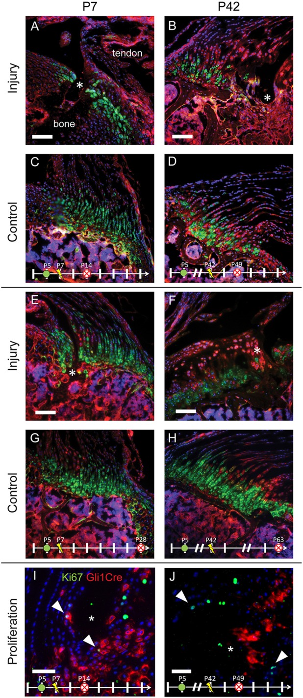
Fig. 2.

The enthesis progenitor population participates in remodeling immature but not mature entheses. Gli1-CreERT2;mTmG mice were injected with TAM on P4 to label the potentially Hh-responsive cell lineage (green) that populates the mature mineralized fibrocartilage. Mice were injured on P7 (A,C,E,G,I) or P42 (B,D,F,H,J) and killed 7 (A-D) or 21 (E-H) days later. Clusters of cells populating the injury regions (*) in A and E are green, indicating that they are part of the original enthesis cell lineage (contralateral controls in C,G). In contrast, cells populating the injury regions (*) in B and F are predominantly red, indicating that they are not derived from the original enthesis cell lineage. To determine the fraction of Gli1 lineage cells that were proliferating, Gli1-CreERT2 mice were crossed with Ai14 mice and stained for Ki67. (I,J) Proliferating cells (green, Ki67) colocalized with Gli1-Cre expression (red) in injured immature entheses (I) but not in mature entheses (J). Arrowheads indicate Ki67+ cells. Scale bars: 100 μm (A-H); 50 μm (I,J). n=5-7 per group. mTmG cells exhibit red fluorescence in the absence of Cre and green fluorescence in presence of Cre. Nuclei are shown in blue. Other colors (e.g. magenta, yellow) are the result of overlapping red, green and/or blue signals.
