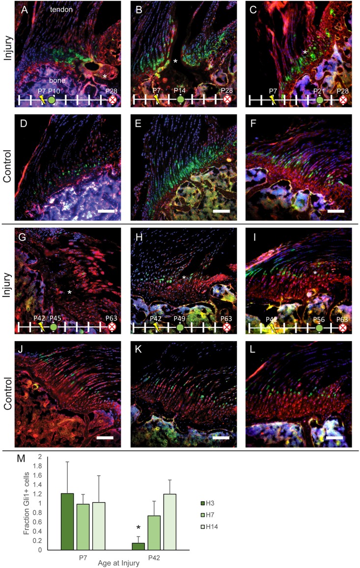
Fig. 3.
Gli1 expression is suppressed in mature healing entheses. (A-F) Entheses of Gli1-CreERT2;mTmG mice injured on P7 (A-C) with matching controls shown below (D-F). (G-L) Entheses of Gli1-CreERT2;mTmG mice injured on P42 (G-I) with matching controls shown below (J-L). TAM was injected at 3 (A,G), 7 (B,H) or 14 days (C,I) after injury to induce labeling of the Gli1+ (green) cell population. Clusters of Gli1+ cells were observed near the defect (*) at all time points after injury in immature entheses (A-C), but few Gli1+ cells were observed 3 days post injury in mature entheses. Scale bars: 100 μm. (M) Quantification of the number of Gli1+ cells in the injured enthesis normalized to that of the Gli1+ cells in contralateral controls after 3 (H3), 7 (H7) and 14 (H14) days of healing. *P<0.05 relative to contralateral controls, two-tailed paired t-test, mean±s.d., n=4-7 per group. mTmG cells exhibit red fluorescence in the absence of Cre and green fluorescence in presence of Cre. Nuclei are shown in blue. Other colors (e.g. magenta, yellow) are the result of overlapping red, green and/or blue signals.

