Fig. 5.
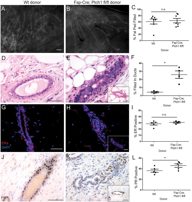
Whole-gland transplantation rescues stunted ducts and ER/PR levels, but not histology, of Fsp-Cre;Ptch1fl/fl animals. Genotype indicates donor glands transplanted to SCID/bg recipients that are wild type for Ptch1. (A,B) Fluorescent whole-mount (A) control and (B) Fsp-Cre;Ptch1fl/fl donor glands, 8 weeks post-transplantation. (C) Quantification of fat pad filling, indicating no difference between groups. (D,E) Hematoxylin and Eosin-stained (D) control and (E) Fsp-Cre;Ptch1fl/fl donor ducts. (F) Quantification of ductal filling, showing increased ductal filling in mutant donors. (G,H) ERα-stained (G) control and (H) Fsp-Cre;Ptch1fl/fl donor ducts. (I) ERα quantification showing similar expression between groups. (J,K) PR-stained (G) control and (H) Fsp-Cre;Ptch1fl/fl donor ducts. (L) PR quantification showing a small increase in Fsp-Cre;Ptch1fl/fl donor ducts. Graphs show data as mean±s.e.m. Insets display histologically normal ducts. *P<0.05 by paired t-test. Insets display histologically normal ducts (E,H,K). n.s., not significant. Scale bars: 0.5 mm in A,B; 50 µm in D,E,G,H,J,K.

