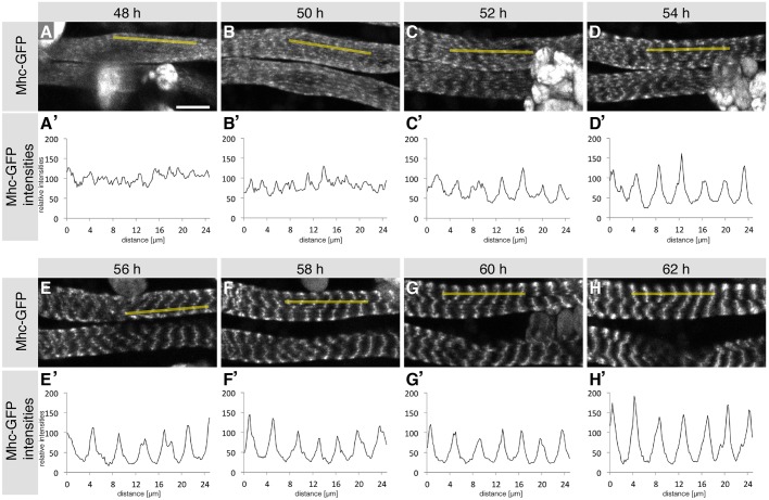
Fig. 2.
Formation of cross-striated abdominal body muscles from live imaging experiments. (A-H) Images of two developing dorsal abdominal muscles expressing Mhc-GFP at 48 h (A), 50 h (B), 52 h (C), 54 h (D), 56 h (E), 58 h (F), 60 h (G) and 62 h (H) APF from a multi-photon movie (Movie 2). (A′-H′) Contrast-adjusted Mhc-GFP intensities within one abdominal dorsal muscle (yellow lines in A-H) at the respective time points. Note the simultaneous appearance of Mhc-GFP periodicity from 50-52 h onwards and its lateral alignment. Scale bar: 10 µm.

