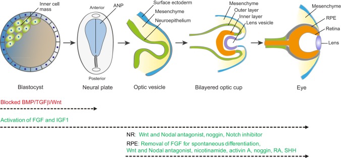
Fig. 1.
Schematic of the key stages of retina development. Beginning with the blastocyst, which contains the pluripotent inner cell mass, gastrulation and neurulation lead to formation of the neural plate. The early eye field is located in the anterior neural plate (ANP) and develops into the optic vesicles. Blocking the activity of BMP, TGFβ and Wnt (red) promotes ANP development. Invagination of the optic vesicle leads to formation of the bilayered optic cup. The inner layer of the optic cup develops into the neural retina (NR) and the outer layer develops into the retinal pigment epithelium (RPE). Activation of FGF and IGF1 pathways (green) facilitates not only development of the ANP but also subsequent optic vesicle/cup formation and retina development. Wnt, FGF, BMP, Notch, SHH, RA and activin A signaling pathways (green) are involved in the specification of the RPE and NR. These factors have been used to promote NR and RPE production from stem cells in vitro, but the specific combinations and concentrations of each factor and the schedule of addition remain to be optimized for each lineage. It is also possible that additional factors not yet identified, and potentially specific to human retinal development, will aid neural retinal and RPE differentiation.

