Fig. 5.

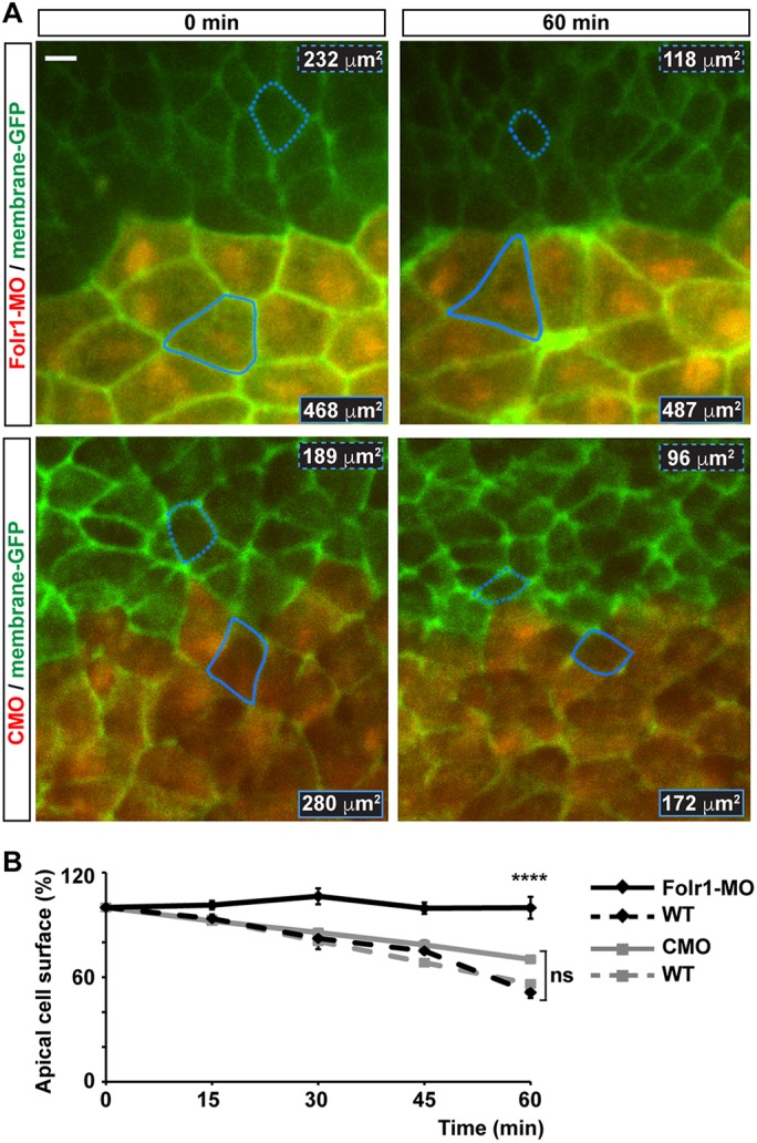
Medial neural plate cells deficient in folate receptor 1 fail to constrict apically during neural plate folding. Two-cell-stage embryos were unilaterally microinjected with 10 pmol morpholino against Folr1 (Folr1-MO) or control morpholino (CMO) along with Alexa Fluor 594-dextran conjugate and bilaterally injected with membrane-GFP. Apical surface of superficial neural plate was time-lapse imaged from whole embryos at a rate of 0.2 min−1. Regions of interest were selected to contour cells close to the midline that remained visible during the 1-h recording and did not divide during this period. (A) Shown are representative examples for the indicated time points of Folr1-MO- and CMO-unilaterally injected and imaged embryos. Outlined is one wild-type (dashed) and one MO-containing (solid) cell for which the apical surface was measured over time. Numbers indicate apical cell surface for the same outlined cells at 0 and 60 min of recording. Scale bar: 10 μm. (B) Graph shows apical cell surface (as percentage of initial cell surface at 0 min, 100%) at the indicated time points. Mean±s.e.m.; n=30 Folr1-MO-, 28 CMO-containing cells, and 30 contralateral wild-type (WT) cells per group; ****P<0.0001; ns, not significant; two-way ANOVA.
