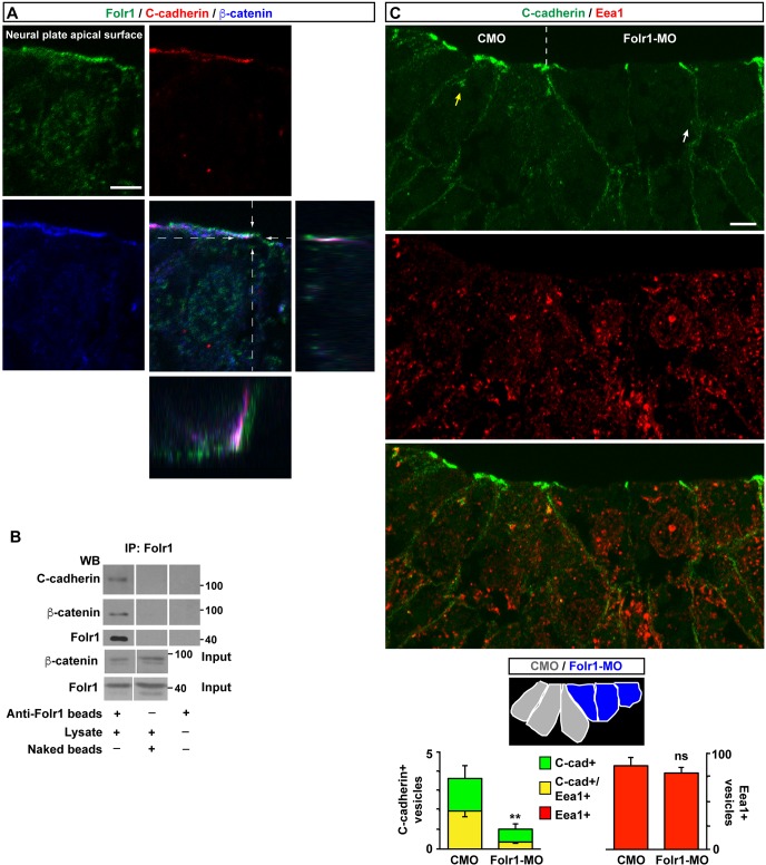
Fig. 6.
Folate receptor interacts with C-cadherin and is necessary for its endocytosis in medial neural plate cells during neural plate folding. (A) Neural plate stage embryos were processed for Folr1 (green), C-cadherin (red) and β-catenin (blue) immunostaining. Shown is a representative transverse single z-section of immunostained stage 15 medial neural plate cell with orthogonal view through the indicated planes to demonstrate partial apical colocalization of Folr1, C-cadherin and β-catenin in the neural plate. Scale bar: 5 µm. (B) C-cadherin and β-catenin co-immunoprecipitate with Folr1. Lysates from wild-type neural plate stage embryos were incubated with anti-Folr1-crosslinked beads or naked beads. Co-immunoprecipitated proteins were dissociated from beads and run in SDS-PAGE gels for western blot assays with anti-Folr1, C-cadherin and β-catenin antibodies. Shown are representative western blot assays, which were performed five times with similar results. (C) The number of C-cadherin-containing vesicles including C-cadherin-containing early endosomes is reduced in Folr1-deficient medial neural plate cells. Two-cell-stage embryos were unilaterally microinjected with CMO and Folr1-MO in contralateral blastomeres. Shown is a representative example of transverse section from a neurulating embryo immunostained for C-cadherin (green) and Eea1 (red). Schematic indicates the cells from which the number of immunopositive vesicles were counted. Yellow arrow points to a C-cadherin+/Eea1+ vesicle and white arrow points to a C-cadherin+/ Eea1− vesicle. Scale bar: 10 µm. Graph shows mean±s.e.m. C-cadherin- and EEA1-labeled vesicles/cell measured in 54 CMO- and 54 Folr1-MO-containing medial neural plate cells in 18 sections from five embryos; **P<0.005; paired Student's t-test.

