Fig. 2.
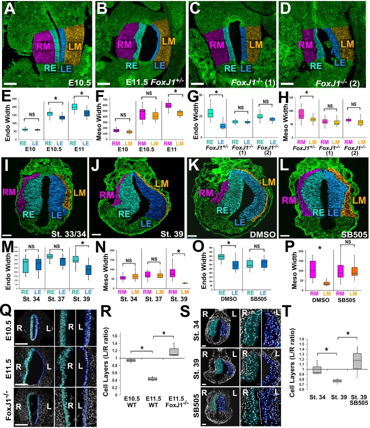
Asymmetries in tissue architecture are regulated by left-right patterning. Sections through mouse (A-D) or frog (I-L) stomachs were stained for β-catenin (A-D) or integrin (I-L) (green) and false color-coded as in Fig. 1 (RE, right endoderm; LE, left endoderm; RM, right mesoderm; LM, left mesoderm). The widths of the endoderm (Endo) and mesoderm (Meso) are significantly different by E10.5-11 in mouse (E,F) and stage 39 in frog (M,N). In Foxj1+/− controls (E11.5), the lumen expands leftward and left-right differences in tissue width are evident (B); however, in Foxj1−/− mutants, the normal leftward expansion of the stomach is perturbed (C,D), and left-right differences in tissue width are eliminated (G,H). Likewise, frog embryos exposed to DMSO show normal leftward expansion of the stomach lumen (K); this is eliminated in embryos exposed to a Nodal inhibitor (SB505124; L), as are normal left-right differences in widths of endoderm and mesoderm (O,P). Nuclear staining reveals asymmetry in the number of endoderm cell layers in the right (teal) versus left (blue) stomach walls by E11.5 in mouse (Q,R) and stage 39 in frog (S,T); this asymmetry is perturbed in Foxj1−/− mutants (Q,R) and in frog embryos exposed to SB505124 (S,T). Scale bars: 100 µm in A-D,Q; 75 µm in I-L,S. *P<0.01; NS, not significant.

