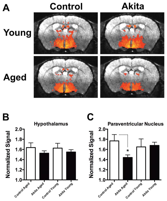
Figure 1. Basal hypothalamic neuronal activity is depressed in 8 month old Ins2Akita mice.
A) Averaged signal intensity maps (MEMRI) plotted on real brain atlas transverse sections in the central hypothalamus (PVN region). Upper row: averaged signal intensity maps from 2 month old Ins2Akita mice (right), and 2 month old controls (left) showing little differences in most regions. Lower row: averaged signal intensity maps from 8 month old Ins2Akita mice (right) and 8 month old controls (left) showing relative hypothalamic and PVN depression in Ins2Akita mice. B) Bar chart showing quantification of normalized signal intensity for the entire hypothalamus and the PVN region (C) in all 4 experimental conditions.

