Figure 1.
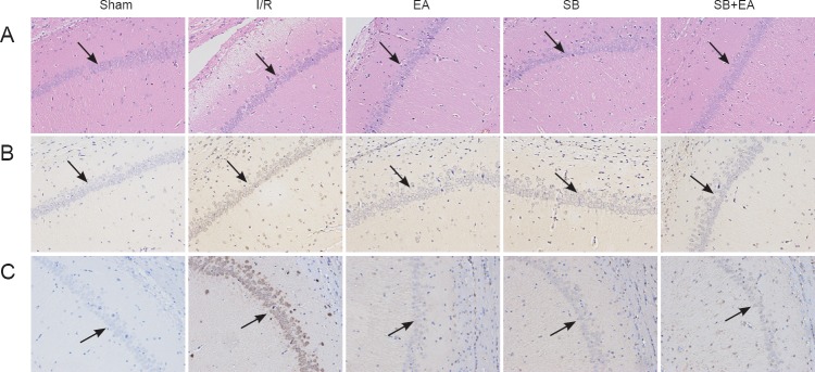
Apoptosis and phosphorylated p38 MAPK-immunoreactive positive cells in the CA1 area of the hippocampus at 3 days after surgery in all groups (light microscope, × 200).
(A) Morphological changes in the hippocampal CA1 area at 3 days after reperfusion. Morphology of cerebral tissues from each group was processed by hematoxylin and eosin staining. (B) Evaluating apoptosis in the hippocampal CA1 area at 1 day after surgery using TUNEL assay. Nuclei of all cells were visualized by DAPI staining, and apoptotic cells were stained brown. (C) Evaluation of phosphorylated p38 MAPK-immunoreactive positive cells in the hippocampal CA1 area at 3 days after surgery using immunohistochemical assay. Sham group: The carotid triangle was exposed, and no line knot was applied; I/R group: cerebral I/R model establishment; EA group: only stimulated by EA and p38 MAPK inhibition; SB group: p38 MAPK blocker SB20358 was injected 30 minutes before model induction; SB + EA group: stimulated by EA and injected with SB20358. I/R: Ischemia/reperfusion; EA: electroacupuncture; SB: p38 MAPK inhibition; MAPK: mitogen-activated protein kinases; DAPI: 4′,6-diamidino-2-phenylindole.

