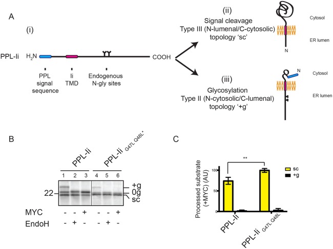
Fig. 6.
Mycolactone sensitivity is dependent upon which TMD-flanking region is translocated. (A) A chimeric protein containing Ii downstream of a pre-prolactin (PPL) signal sequence (i) and the two topologies it might assume following integration into RMs, depending on whether the region that is translocated is N-terminal (ii) or C-terminal (iii) of the TMD. (B) Translation of PPL-Ii and PPL-IiG47L Q48L* in the absence or presence of mycolactone (MYC), followed by treatment with EndoglycosidaseH (EndoH). Samples were analysed following immunoprecipitation of Ii. (C) Graph showing the amount of signal-cleaved (‘sc’) or glycosylated (‘+g’) substrate in the presence of mycolactone relative to control samples. These values were determined by dividing the quantity of ‘sc’ or ‘+g’ substrate obtained in the presence of mycolactone by the quantity of ‘sc’ or ‘+g’ substrate obtained in the absence of mycolactone and are expressed as percentages. The statistical test performed was two-way ANOVA. Error bars show mean±s.d. (n=3). P-values and other symbols are as defined in Fig. 1 legend.

