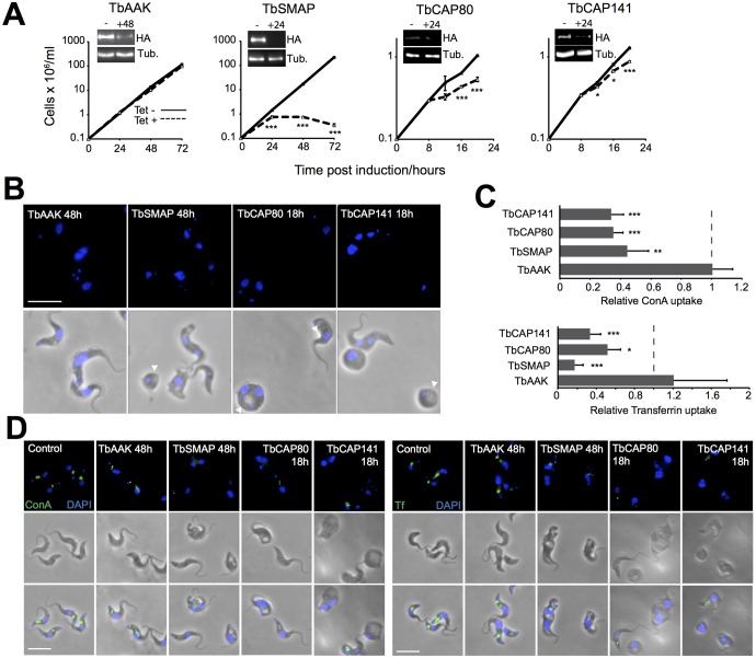
Fig. 5.
Phenotypic consequences of clathrin-associated protein depletion. Tetracycline-inducible RNAi cell lines for selected clathrin-associated proteins were generated in HA-tagged bloodstream-form cells. (A) Effects of tetracycline addition on protein levels as assessed by western blotting before RNAi induction and 24 h after induction using an anti-HA antibody (insets, - and +24, respectively), and its effect on cell proliferation expressed as mean±s.e.m. from three independent repeats. – (dashed lines), uninduced controls. (B) Morphology of induced RNAi cells at the indicated time points. Blue, DAPI staining; white arrowheads denote vacuolar structures viewed under phase-contrast conditions that are reminiscent of swollen flagellar pockets. (C,D) Effects of clathrin-associated protein depletion on uptake of FITC–transferrin or FITC–ConA. (C) Quantification of FITC–transferrin or FITC–ConA uptake in induced RNAi cells versus uninduced controls. Data are mean±s.d. of at least 50 cells per condition from two independent experiments normalised to non-induced controls. (D) Representative images showing FITC–transferrin or FITC–ConA (green) accumulation following a 45-min pulse. Blue, DAPI staining. Scale bars: 5 μm. *P<0.05, **P<0.005, ***P<0.001 versus control (t-test).

