Fig. 6.
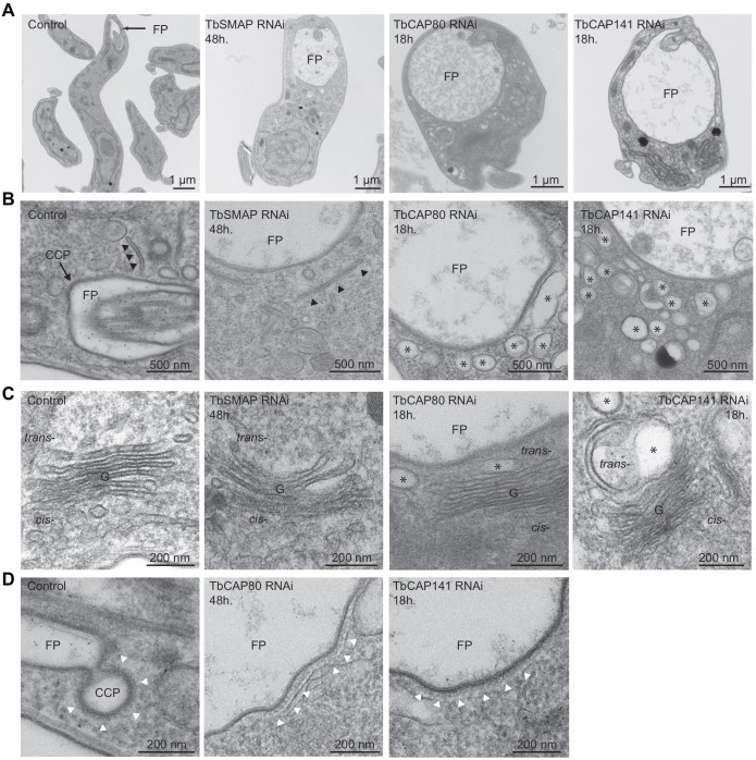
Effects of clathrin-associated protein depletion on endomembrane system ultrastructure. Representative transmission electron micrographs of ultrathin resin sections of bloodstream-form parasites after RNAi-mediated depletion of clathrin-associated proteins. (A) Gross ultrastructural defects, in particular swelling of the flagellar pocket (FP) and cytoplasmic vacuolisation. (B) Tubular endosomes (black arrowheads) are apparent in the vicinity of the flagellar pocket of control and TbSMAP-depleted cells. A clathrin-coated pit (CCP) profile is seen at the flagellar pocket membrane of the control section (black arrowheads). Extensive vacuolisation (asterisks) is apparent in both TbCAP80- and TbCAP141-depleted cells. (C) Golgi (G) profiles. The control cell shows numerous ER–Golgi transport intermediates. TbSMAP depletion has little apparent effect upon Golgi ultrastructure, whereas depletion of TbCAP80 or TbCAP141 causes vacuolisation and swelling (asterisks) of the trans-cisternae. (D) Clathrin-coated profiles (white arrowheads) of control and TbCAP80- or TbCAP141-depleted cells. Aberrant large and flat coated profiles are seen in TbCAP80- or TbCAP141-depleted cells.

