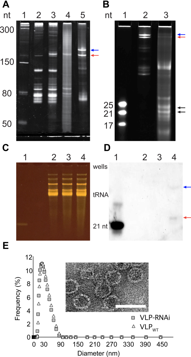
Figure 2.
Characterization of the RNAi scaffold and VLP–RNAi by electrophoresis, microscopy, Northern blotting and dynamic light scattering. (A) The in vivo transcribed RNAi scaffold was produced in E. coli. Lane 1: Size ladder. Lane 2: Total E. coli RNA before induction of expression of the RNAi scaffold and CP. Lane 3: Total E. coli RNA after induction. Lane 4: RNA extracted from purified VLP–RNAi. Lane 5: RNAi scaffold prepared by in vitro transcription. The red arrow indicates the RNAi scaffold. The blue arrow indicates RNAi scaffold extended by read-through of the terminator. (B) RNAi scaffold prepared by in vitro transcription and then processed by human Dicer. Lane 1: Size ladder. Lane 2: RNAi scaffold. Lane 3: RNAi scaffold treated with human Dicer. The black arrows indicate products of Dicer processing of the RNAi scaffold. The Dicer products are 21- to 25-mers. Panels C and D show an assay for products of in vivo processing of VLP–RNAiLet-7 in U87 cells. (C) All small RNAs visualized on a denaturing gel. (D) The same gel with the processed RNAiLet-7 scaffold visualized by Northern blotting with an antisense DNA probe. Lane 1: The 21 nt Let-7 siRNA is a positive control. Lane 2: RNA from cells incubated with PBS are a negative control. Lane 3: RNA from cells incubated with VLPWT are a negative control. Lane 4: RNA from cells incubated with VLP–RNAiLet-7. The red arrow indicates product of in vivo Dicer processing. The blue arrow indicates incomplete cleavage by Dicer. Six hundred nanograms of enriched small RNA were loaded in each lane, except for the positive control, which is 50 ng of RNA. (E) Characterization of Qβ-VLP with RNAi scaffold (gray rectangle) and wild-type Qβ-VLP (white triangle) by dynamic light scattering. Inset: Transmission electron microscope images of Qβ VLP containing the RNAi scaffold. Scale bar = 50 nm. The gels are denaturing.

