Fig. 4.

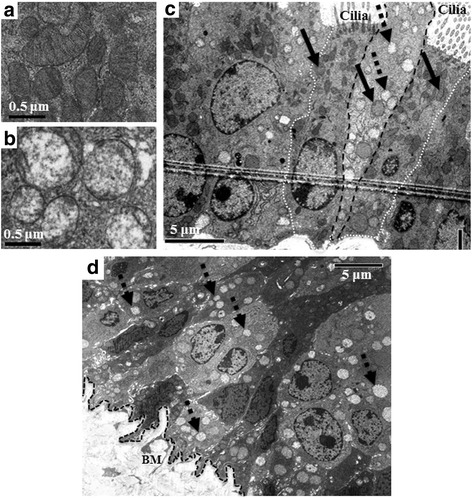
Detection of cells with a high number of morphologically abnormal mitochondria. Sections from WT, Aldh2*2 Tg, and Aldh2 −/− adult mice as well as from aged WT mice were examined with TEM for the presence of cellular ultrastructure abnormality. Compared to the typical shaped normal mitochondria (a), morphologically abnormal mitochondria, with rounded shape, complete lack of cristae, and a marked decrease in matrix density were detected in all groups (b). Some cells showed most mitochondria of the abnormal type (dotted arrows) and a few mitochondria of the normal type (solid arrows) in club (dashed black line) and basal cells of all mice, but not in their ciliated cells (dotted white line) (c) (a, b, and c are representative photos from WT). Aldh2 −/− tracheas showed a large number of cells with >80% mitochondria being abnormal (dotted arrows) (d). BM: Basement membrane
