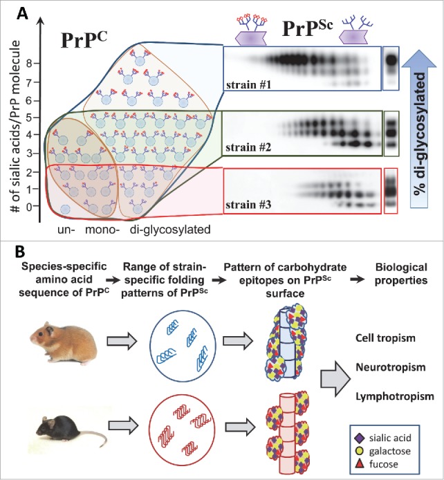
FIGURE 2.

(A) Schematic diagram illustrating that PrPSc strains recruit PrPC isoforms selectively according to PrPC glycosylation and sialylation status (adopted from12). Strain #1 recruits sialoglycoforms of PrPC without noticeable preferences. Hypersialylated and diglycosylated PrPC are preferentially excluded from the strain #2 and even more so from the strain #3. As a result, the proportion of hyper- versus hyposialylated molecules within PrPSc (illustrated by the 2D Western blots), as well as the ratios of di-, mono- and unglycosylated glycoforms (shown on 1D Western blots on right hand side), changes in a strain-specific manner. PrPC molecules are shown as blue circles and sialic acid residues - as red diamonds. (B) Schematic diagram illustrates that species-specific amino acid sequences of PrPC determine the range of strain-specific folding patterns, which define the strain-specific patterns of functional carbohydrate epitopes on PrPSc surfaces within a particular host. Patterns of functional carbohydrate epitopes determine strain-specific biologic properties. PrPSc folding patterns are expected to be quite different for 2 groups of strains: (i) a group that can accommodate diglycosylated glycoforms and (ii) another group that selectively excludes diglycosylated glycoforms.
