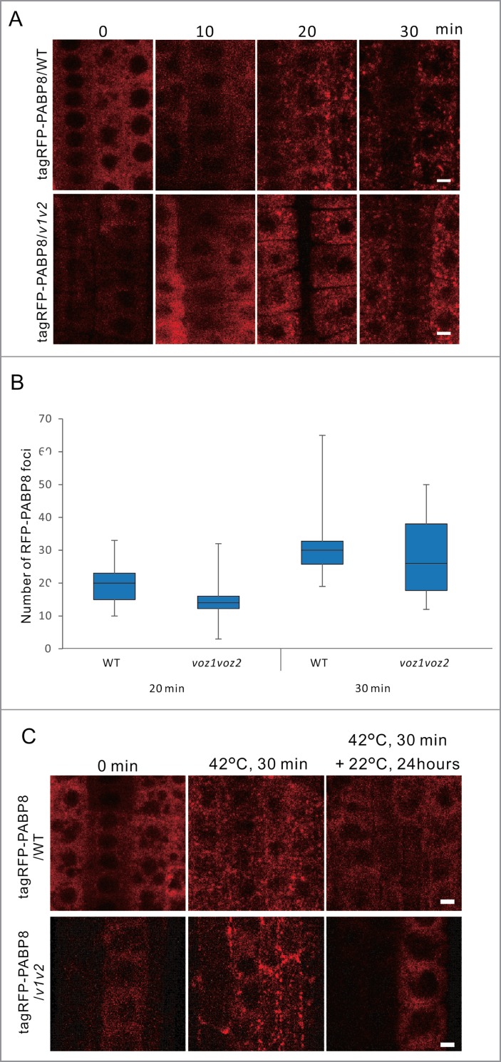
Figure 4.
The granule structures of PABP8 in WT or the voz1voz2 mutant under heat stress conditions. (A) Subcellular localization of tagRFP-PABP8 was observed in 7-day-old WT and voz1voz2 seedlings. (B) The box-and-whisker plot shows the number of tagRFP-PABP8 foci (SG markers) in root tip cells of WT and voz1voz2 lines were treated at 42 °C for 20 min or 30 min. P values were calculated by the Mann–Whitney U test (*P < 0.05). (C) Seven-day-old WT and voz1voz2 seedlings expressing tagRFP-PABP8 were incubated at 42 °C for 30 min, followed by incubation at 22 °C for 24 h, and images of root tip cells were captured using the confocal microscope. Scale bars = 5 µm.

