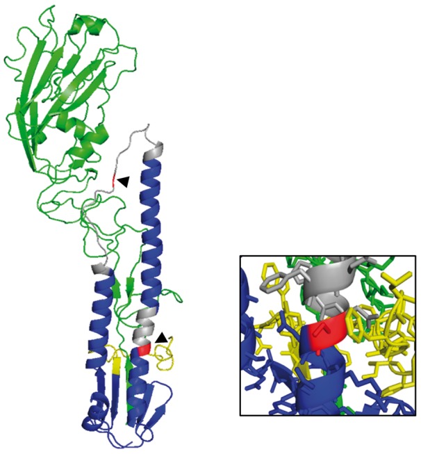
Figure 5.

Schematic illustration of the situation of the two mutations reported here within the H1 hemagglutinin structure for wild duck strain WDK/JX/12416/2005. The mutated positions are shown in red and indicated by arrowheads. Green indicates the HA1 subunit, blue indicates the HA2 subunit with the fusion peptide in yellow and regions changing conformation at acidic pH to trigger fusion in grey. The insert shows a close up of the HA2 residue 113 region with side chains displayed (the wild-type serine 113 is shown). Residue 113 is close to a phenylalanine side chain at position 3 of the fusion peptide. The structure was generated based on Protein Data Bank file accession number 3HTO (Lin et al. 2009) using the PyMOL molecular graphics system version 1.7.5.0.
